
form of the head and neck – pdf
The head and neck region is a complex anatomical structure, blending aesthetic and functional roles․ Its intricate design supports vital functions like respiration, digestion, and sensory perception, making it a focal point for both medical professionals and students․ Understanding its detailed anatomy, including cranial bones, facial structures, and musculature, is essential for clinical and surgical applications․ This section provides a foundational overview, highlighting key anatomical landmarks and their significance in human physiology․
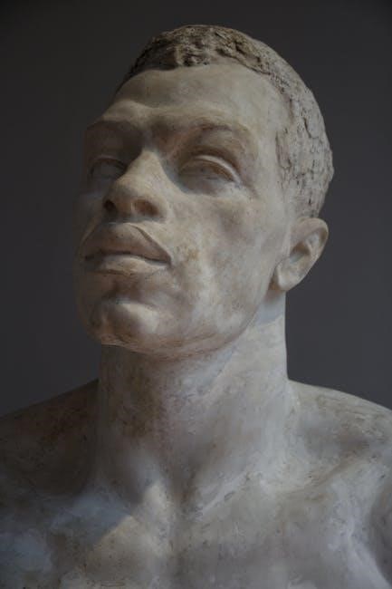
1․1 Overview of the Head and Neck Region
The head and neck region is a complex anatomical area combining aesthetic and functional elements․ It includes cranial bones, facial structures, and cervical compartments, with muscles, nerves, and blood vessels intricately organized․ This region supports vital functions like respiration, digestion, and sensory perception․ Its musculo-aponeurotic layer and cervical plexus are key components, making it a critical area for clinical and surgical studies․

1․2 Importance of Studying Head and Neck Anatomy
Studying head and neck anatomy is crucial for understanding cranial nerves, lymphatic drainage, and musculo-aponeurotic layers․ It aids in diagnosing conditions like trigeminal neuralgia and planning surgeries․ Knowledge of cervical plexus and fascia is vital for clinical correlations․ This study also enhances comprehension of pharyngeal structures and oral cavity subdivisions, essential for dentistry and otolaryngology․ It bridges anatomy with clinical practice, improving patient outcomes․

Bones and Joints of the Head and Neck
The head and neck contain cranial bones, facial bones, and the mandible, forming a structural framework․ The temporomandibular joint (TMJ) facilitates jaw movement, while bones support sensory organs, nerves, and blood vessels․
2․1 Cranial Bones (Neurocranium)
The cranial bones form the neurocranium, which protects the brain and houses sensory organs․ In adults, these bones are fused, creating a rigid structure․ The frontal, occipital, and sphenoid bones are key components, anchoring muscles and forming the cranial cavity․ They also support the facial bones and contribute to the skull’s structural integrity, ensuring proper alignment and function of the head and neck region;
2․2 Facial Bones and Mandible
The facial bones, including the maxilla, zygoma, and mandible, form the structural framework of the face․ The mandible, or lower jawbone, articulates with the skull via the temporomandibular joint (TMJ)․ These bones support facial features, enable mastication, and protect vital structures․ Their intricate design allows for both aesthetic appeal and functional efficiency, making them critical in head and neck anatomy studies and clinical applications․
2․3 Temporomandibular Joint (TMJ)
The temporomandibular joint (TMJ) connects the mandible to the skull, enabling jaw movements essential for chewing, speaking, and yawning․ It is a synovial joint with articular discs that facilitate smooth articulation․ The TMJ is vital for mastication and maintaining facial symmetry, making it a key focus in both anatomical studies and clinical applications for treating disorders like temporomandibular dysfunction․
Cranial Nerves and Their Functions
Cranial nerves are 12 pairs of nerves originating from the brain, controlling functions like sensation, movement, and involuntary actions․ They include the trigeminal, facial, and vagus nerves, each with specialized roles in sensory input, motor control, and autonomic functions, making them critical for head and neck physiology and clinical diagnostics․
3․1 Trigeminal Nerve (CN V)
The trigeminal nerve (CN V) is the fifth cranial nerve, divided into three branches: ophthalmic, maxillary, and mandibular․ It provides sensory innervation to the face, eyes, and oral cavity while controlling muscles like the temporalis and masseter for mastication․ Its mandibular division also supplies the lower face and jaw, playing a key role in chewing and facial sensation, with clinical relevance in conditions like trigeminal neuralgia․
3․2 Facial Nerve (CN VII) and Its Role
The facial nerve (CN VII) is the seventh cranial nerve, responsible for controlling muscles of facial expression, taste sensation in the anterior tongue, and providing parasympathetic innervation to salivary and tear glands․ It also regulates the stapedius muscle in the middle ear․ Damage to CN VII can lead to conditions like Bell’s palsy, affecting facial symmetry and emotional expression, underscoring its vital role in both motor and sensory functions;
3․3 Other Cranial Nerves in the Head and Neck
Beyond CN V and CN VII, other cranial nerves in the head and neck include the glossopharyngeal (CN IX), vagus (CN X), accessory (CN XI), and hypoglossal (CN XII) nerves․ CN IX manages taste, salivation, and swallowing, while CN X regulates visceral functions, heart rate, and digestion․ CN XI controls neck muscles, and CN XII governs tongue movements, each playing distinct roles in sensory and motor functions․

Fascia and Cervical Plexus
The head and neck region is organized by fascial layers, separating muscles and organs․ The cervical plexus, a network of nerves, innervates neck muscles and provides sensory supply․
4․1 Musculo-Aponeurotic Layer of the Head and Neck
The musculo-aponeurotic layer consists of broad, fibrous sheets and muscles, such as the occipital and frontal bellies․ It provides structural support and facilitates movements of the head and neck․
4․2 Visceral and Vascular Compartments of the Neck
The neck is organized into four compartments: visceral, vertebral, and two vascular․ The visceral compartment contains essential organs like the pharynx, larynx, and trachea, while the vascular compartment houses major blood vessels, including the carotid arteries and jugular veins․ These compartments are crucial for maintaining vital functions and structural integrity, aiding in diagnosis and surgical interventions․
Blood Supply and Venous Drainage
The head and neck receive blood supply from the common carotid and vertebral arteries, branching into external and internal carotid systems․ Venous drainage is primarily via jugular veins, ensuring efficient blood circulation and clinical significance in surgical procedures․
5․1 Arteries of the Head and Neck
The head and neck are primarily supplied by the common carotid and vertebral arteries, branching into external and internal carotid systems․ These arteries provide blood to facial structures, scalp, and intracranial regions․ The external carotid artery supplies external structures, while the internal carotid artery serves the brain and eyes․ This network ensures vital oxygenation and nutrient delivery to the complex tissues of the head and neck․
5;2 Veins of the Head and Neck
The venous system of the head and neck is a dual system, comprising superficial and deep veins․ Superficial veins, like the jugular and facial veins, drain surface structures, while deep veins, such as the sigmoid sinus, drain intracranial and orbital regions․ The external and internal jugular veins serve as major pathways for venous blood drainage, ensuring proper circulation and maintaining the region’s physiological balance․

Lymphatic Drainage of the Head and Neck
The lymphatic system of the head and neck plays a crucial role in immune function and detoxification․ It comprises lymph nodes distributed in specific regions, aiding in the filtration of lymph and detection of pathogens․ This network is vital for maintaining health and is often a focus in clinical diagnostics and treatments․
6․1 Lymph Nodes and Their Distribution
The head and neck contain numerous lymph nodes, primarily located in the cervical, submandibular, and parotid regions․ These nodes play a vital role in immune defense by filtering lymph and detecting pathogens․ Their strategic distribution facilitates the drainage of lymph from various tissues, ensuring efficient immune response․ Understanding their distribution is crucial for clinical diagnostics and treatments, particularly in managing infections and cancers․
6․2 Clinical Significance of Lymphatic Drainage
Lymphatic drainage in the head and neck is crucial for detecting and managing diseases like cancer․ It aids in identifying metastasis and planning targeted therapies․ Proper drainage prevents infections and inflammation, ensuring tissue health․ Clinicians rely on lymph node examinations to diagnose conditions, making this system vital for both diagnostic and therapeutic interventions in head and neck pathology․

Oral Cavity and Pharynx Anatomy
The oral cavity includes structures like lips, tongue, and buccal mucosa, while the pharynx facilitates swallowing and speech․ Understanding their anatomy is crucial for diagnosing oral pathologies and planning treatments, emphasizing the interconnected roles of these regions in digestion, respiration, and communication․
7․1 Anatomic Subdivisions of the Oral Cavity
The oral cavity is divided into the lips, floor of the mouth, oral tongue (anterior two-thirds), buccal mucosa, upper and lower gingivae, hard palate, and retromolar trigone․ These subdivisions play critical roles in mastication, speech, and deglutition․ Their distinct anatomical features and functions are essential for understanding oral pathologies and surgical interventions, as detailed in Vishram Singh’s textbook and other anatomical resources․

7;2 Pharyngeal Structure and Function
The pharynx is divided into three regions: nasopharynx, oropharynx, and laryngopharynx․ It plays a crucial role in swallowing and airway protection․ The nasopharynx connects with the nasal cavity, while the oropharynx and laryngopharynx facilitate food passage to the esophagus․ Pharyngeal muscles, including the pharyngeal constrictors, coordinate with the epiglottis to prevent aspiration, ensuring efficient digestion and respiration, as detailed in head and neck anatomy resources․

Clinical and Surgical Landmarks
The posterior triangle of the neck is bounded by the sternocleidomastoid, trapezius, and omoclavicular triangle, serving as key landmarks for surgical interventions and clinical examinations, as detailed in anatomy resources․
8․1 Boundaries of the Posterior Triangle of the Neck
The posterior triangle of the neck is bordered by the sternocleidomastoid muscle anteriorly, the trapezius muscle posteriorly, and the omoclavicular triangle inferiorly․ The clavicle and scapula form additional structural boundaries, creating a region divided into supraclavicular and infraclavicular areas․ These anatomical landmarks are crucial for identifying surgical access points and understanding regional lymphatic drainage patterns, as detailed in head and neck anatomy resources․
8․2 Surgical Implications of Head and Neck Anatomy
Understanding head and neck anatomy is critical for surgical planning and execution․ The complex interplay of nerves, blood vessels, and fascial layers demands precise knowledge to avoid complications․ Surgical landmarks like the posterior triangle boundaries guide procedures, while detailed anatomical maps, such as those in Vishram Singh’s textbook, provide essential references․ This knowledge ensures safe and effective surgical outcomes in the region․
Recommended Resources and PDFs
Vishram Singh’s Textbook of Anatomy (Head, Neck, and Brain) is a comprehensive guide, offering detailed insights and clinical correlations․ Other reliable PDFs provide supplementary material for in-depth study․
9․1 Vishram Singh’s Textbook of Anatomy (Head, Neck, and Brain)
Vishram Singh’s Textbook of Anatomy is a comprehensive guide covering the detailed anatomy of the head, neck, and brain․ It includes clinical correlations and case studies, making it invaluable for medical students and professionals․ The high-quality PDF is available via a Google Drive link, offering insights into cranial bones, facial structures, and muscles․ This resource is essential for understanding the complex anatomy of the head and neck region․
9․2 Other Reliable PDF Resources for Head and Neck Anatomy
Beyond Vishram Singh, other reliable PDF resources include the University of Utah’s lecture syllabus, covering fascia, cervical plexus, and lymphatic drainage․ The Sobotta Atlas of Human Anatomy provides detailed illustrations of the head, neck, and upper limb․ Additionally, BD Chaurasia’s Anatomy offers comprehensive insights into gross anatomy and clinical correlations, making these resources indispensable for in-depth study․